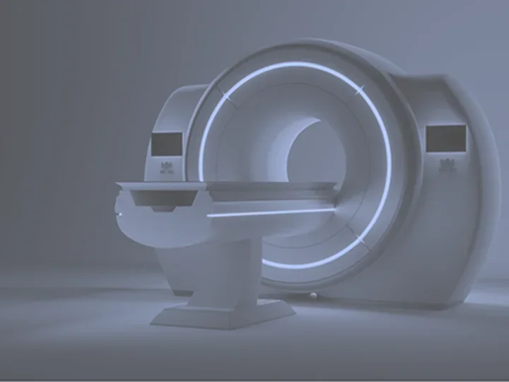
Preclinical MRI (PMRI) Lab
What is the Preclinical MRI (PMRI) lab?
Located in the UCLA Center for Health Sciences, the PMRI is a state-of-the-art, preclinical imaging center open to all types of investigators.
The PMRI employs advanced imaging technologies to support preclinical research and education throughout the UCLA Health campus. Our team of experts physicians, scientists, fellows, and technologists are available to collaborate with investigators worldwide.
Our purpose
The PMRI supports UCLA Health and the broader scientific community in advancing high-quality preclinical research and education. Our mission is to:
- Facilitate and contribute to preclinical studies that prioritize ethical standards, safety, and the humane care of approved and regulated laboratory animal models.
- Support the development and assessment of innovative therapies, technologies, and medical devices.
- Enhance educational and training opportunities for staff, students, and researchers across diverse fields in medicine and science.
Our facilities and equipment
The PMRI has a Bruker BioSpec 3T, a multipurpose high field MR scanner for magnetic resonance imaging (MRI) and magnetic resonance spectroscopy (MRS) for preclinical research and material science applications. The instrument consists of:
- 3 Tesla Magnet with Maxwell Technology, Ultra-Shielded & Refrigerated, with Long Hold Time
- Faraday RF-Shielding Cabinet (CCM)
- Heat Exchanger for Magnet Refrigerator and Gradient/Shim Cooling
- High Power Gradient Amplifier
- Two 1H Parallel Transmit and Receive RF Channels
- Mouse Body Volume RF Coil – 40 mm
- Mouse Head Volume Coil – 23 mm
- Multi-Purpose Planar Surface Coils – 10mm and 20mm
- Mouse Brain Array Coil – 4 Channels
- Motorized Animal Transport System
- Closed Cradle for Small and Large Mice
- Body Temperature Conditioning System
- Warm Water Heating Cover Set for Mice/Rats (for the Motorized Animal Transport System)
- Air Supply & Gas Removal Pump for Multimodal Animal Cradles
- ParaVision® 360 MRI Comprehensive Workplace
Our expertise
The PMRI is supported by a dedicated staff with decades of experience in collaborative research and innovation.
Our services
The PMRI lab specializes in preclinical studies utilizing approved small-animal research models. Our experienced staff work closely with your product development team to provide customized scientific and technical support at every stage of your project.
Our facilities and advanced imaging capabilities support a wide range of applications.
🧠 Imaging Services
High-resolution, noninvasive MRI imaging of small animals (commonly mice and rats) for anatomical, functional, and molecular studies:
- Anatomical imaging (T1-, T2-, and proton density-weighted scans)
- Functional MRI (fMRI) for brain activity mapping
- Diffusion tensor imaging (DTI) for white matter tractography
- Perfusion imaging (arterial spin labeling, dynamic susceptibility contrast)
- Dynamic contrast-enhanced (DCE) MRI for tumor vascularity and permeability studies
- Longitudinal imaging to monitor disease progression or treatment response
🧬 Preclinical Study Support
- Study design and protocol development assistance (animal model selection, imaging schedules, contrast agent selection)
- Animal preparation and monitoring (anesthesia, physiological monitoring, temperature regulation)
- Image acquisition and optimization (custom pulse sequences, parameter tuning)
💻 Image Processing and Quantitative Analysis
- Segmentation, 3D reconstruction, and volumetric analysis
- Tumor or lesion volume quantification
- Signal intensity and relaxation time (T1/T2) mapping
- Parameter extraction
- Statistical analysis and report generation
- Integration with histopathology or molecular endpoints
🧫 Applications and Research Areas
- Neuroscience: brain connectivity, stroke, neurodegeneration, injury models
- Oncology: tumor detection, growth tracking, treatment response, vascularity
- Musculoskeletal imaging: bone, cartilage, and soft tissue evaluation
- Metabolic disease: liver fat quantification, obesity, diabetes models
🧑🔬 Training and Collaboration
- Hands-on training in small animal MRI operation and analysis
- Method development collaborations (sequence optimization, new biomarkers)
- Consultation for grant proposals or study design
🧩 Facility Capabilities
- Dedicated Bruker MRI scanner
- Animal handling suite with anesthesia and monitoring equipment
- Contrast agent preparation and injection setup
- Data storage and analysis workstations
