Clinical Research
In the clinical research domain at the UCLA Brain Tumor Imaging Laboratory (BTIL), our focus is on translating cutting-edge medical imaging technologies into tangible benefits for patients. We are dedicated to advancing diagnostic accuracy, treatment assessment, and patient care through the meticulous analysis of real-world clinical data. By leveraging innovative imaging techniques and applying rigorous quantitative methodologies, our clinical research endeavors aim to improve the understanding and management of brain tumors, ultimately contributing to better outcomes and quality of life for those affected by these conditions.
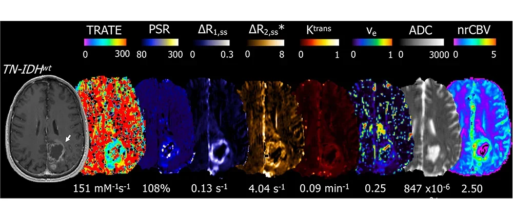
Brain Tumors
Our clinical research is dedicated to unraveling the complexities of brain tumors, encompassing a range of types from gliomas to metastatic lesions centering around the exploration and application of advanced MRI (Magnetic Resonance Imaging) and PET (Positron Emission Tomography) techniques for a comprehensive understanding of brain tumors. Through advanced MRI and PET techniques, researchers aim to:
- Treatment Evaluation: BTIL is committed to assessing the effectiveness of brain tumor treatments. By utilizing advanced imaging, researchers can track changes in tumor size, composition, and metabolic activity, providing vital insights into treatment response and guiding clinical decisions.
- Characterization: The lab specializes in decoding the complex makeup of brain tumors. By employing sophisticated imaging techniques, researchers can discern unique tumor characteristics, such as genetic markers, cellular density, and molecular signatures, aiding in accurate diagnosis and tailored treatment strategies.
- Visualization: Advanced imaging enables BTIL to create detailed visual representations of brain tumors. These visuals enhance the understanding of tumor location, size, and interaction with surrounding brain structures, assisting surgeons and oncologists in planning precise interventions.
- Quantification: Quantitative analysis is a cornerstone of BTIL's research. Through precise measurement and quantification of tumor features, such as blood flow, diffusion, and metabolic activity, researchers can extract quantitative data to inform clinical decisions and monitor treatment progress.
Our goal is to contribute to the advancement of brain tumor research and patient care. The lab's dedication to refining imaging technologies leads to improved diagnostic accuracy, personalized treatment strategies, and enhanced outcomes for individuals affected by brain tumors.
Selected References
Imaging biomarkers for antiangiogenic therapy in malignant gliomas.
Leu K, Pope WB, Cloughesy TF, Lai A, Nghiemphu PL, Chen W, Liau LM, Ellingson BM.CNS Oncol. 2013 Jan;2(1):33-47. doi: 10.2217/cns.12.29.
Ellingson BM, Patel K, Wang C, Raymond C, Brenner A, de Groot JF, Butowski NA, Zach L, Campian JL, Schlossman J, Rizvi S, Cohen YC, Lowenton-Spier N, Minei TR, Shmueli SF, Wen PY, Cloughesy TF. Neurooncol Adv. 2021 Jun 19;3(1):vdab082. doi: 10.1093/noajnl/vdab082.
Ellingson BM, Gerstner ER, Smits M, Huang RY, Colen R, Abrey LE, Aftab DT, Schwab GM, Hessel C, Harris RJ, Chakhoyan A, Gahrmann R, Pope WB, Leu K, Raymond C, Woodworth DC, de Groot J, Wen PY, Batchelor TT, van den Bent MJ, Cloughesy TF. Clin Cancer Res. 2017 Oct 1;23(19):5745-5756. doi: 10.1158/1078-0432.CCR-16-2844.
Neurological, neurodegenerative, and neuropsychiatric diseases
UCLA Brain Tumor Imaging Laboratory (BTIL) specializes in multimodal image processing and analysis, particularly concerning neurodegenerative diseases.
This comprehensive approach enables BTIL to provide a more holistic understanding of neurodegenerative diseases, such as Alzheimer's, Parkinson's, or Huntington's disease and CSM, Schizophrenia, and Epilepsy. By deciphering the combined information from multiple imaging modalities, BTIL contributes to improved disease diagnosis, progression tracking, and treatment evaluation, ultimately leading to better insights into disease mechanisms and more effective strategies for early detection and intervention.
Selected References
Ellingson BM, Gerstner ER, Smits M, Huang RY, Colen R, Abrey LE, Aftab DT, Schwab GM, Hessel C, Harris RJ, Chakhoyan A, Gahrmann R, Pope WB, Leu K, Raymond C, Woodworth DC, de Groot J, Wen PY, Batchelor TT, van den Bent MJ, Cloughesy TF. Clin Cancer Res. 2017 Oct 1;23(19):5745-5756. doi: 10.1158/1078-0432.CCR-16-2844.
Wang C, Ellingson BM, Oughourlian TC, Salamon N, Holly LT. EBioMedicine. 2022 Oct;84:104255. doi: 10.1016/j.ebiom.2022.104255.
Wang C, Oughourlian T, Tishler TA, Anwar F, Raymond C, Pham AD, Perschon A, Villablanca JP, Ventura J, Subotnik KL, Nuechterlein KH, Ellingson BM. Schizophr Res. 2021 May;231:179-188. doi: 10.1016/j.schres.2021.04.001.
Wang C, Tishler TA, Nuechterlein KH, Ellingson BM. Psychiatry Res. 2023 Aug;326:115364. doi: 10.1016/j.psychres.2023.115364.