At the UCLA Brain Tumor Imaging Laboratory, we are pioneers in the development of new imaging techniques and methods to improve the diagnosis, staging, treatment, and monitoring of brain tumors and neurodegenerative diseases. We are committed to translating our research findings into clinical practice to improve the lives of our patients.
Explore our research
Clinical
Brain Tumors
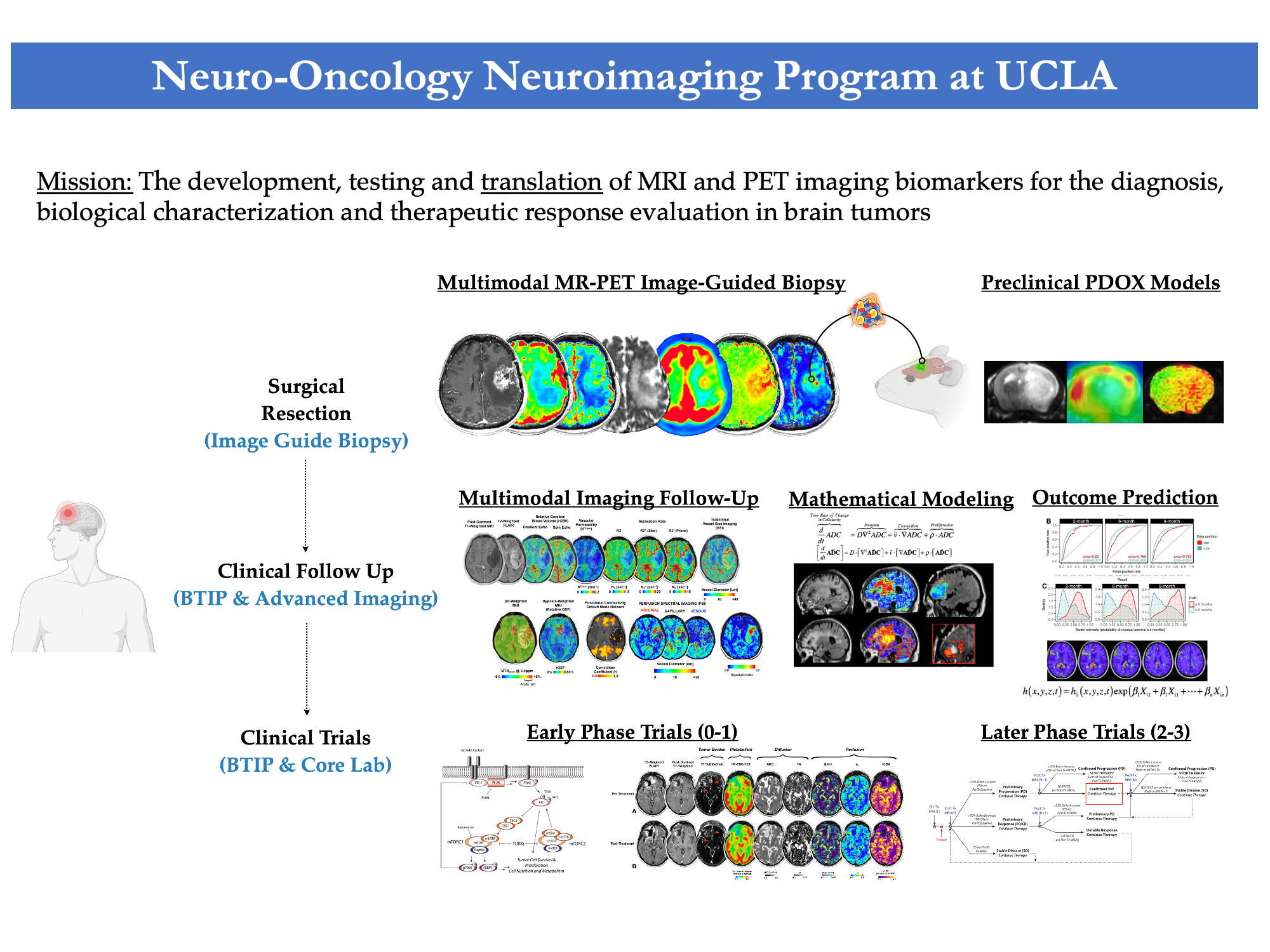
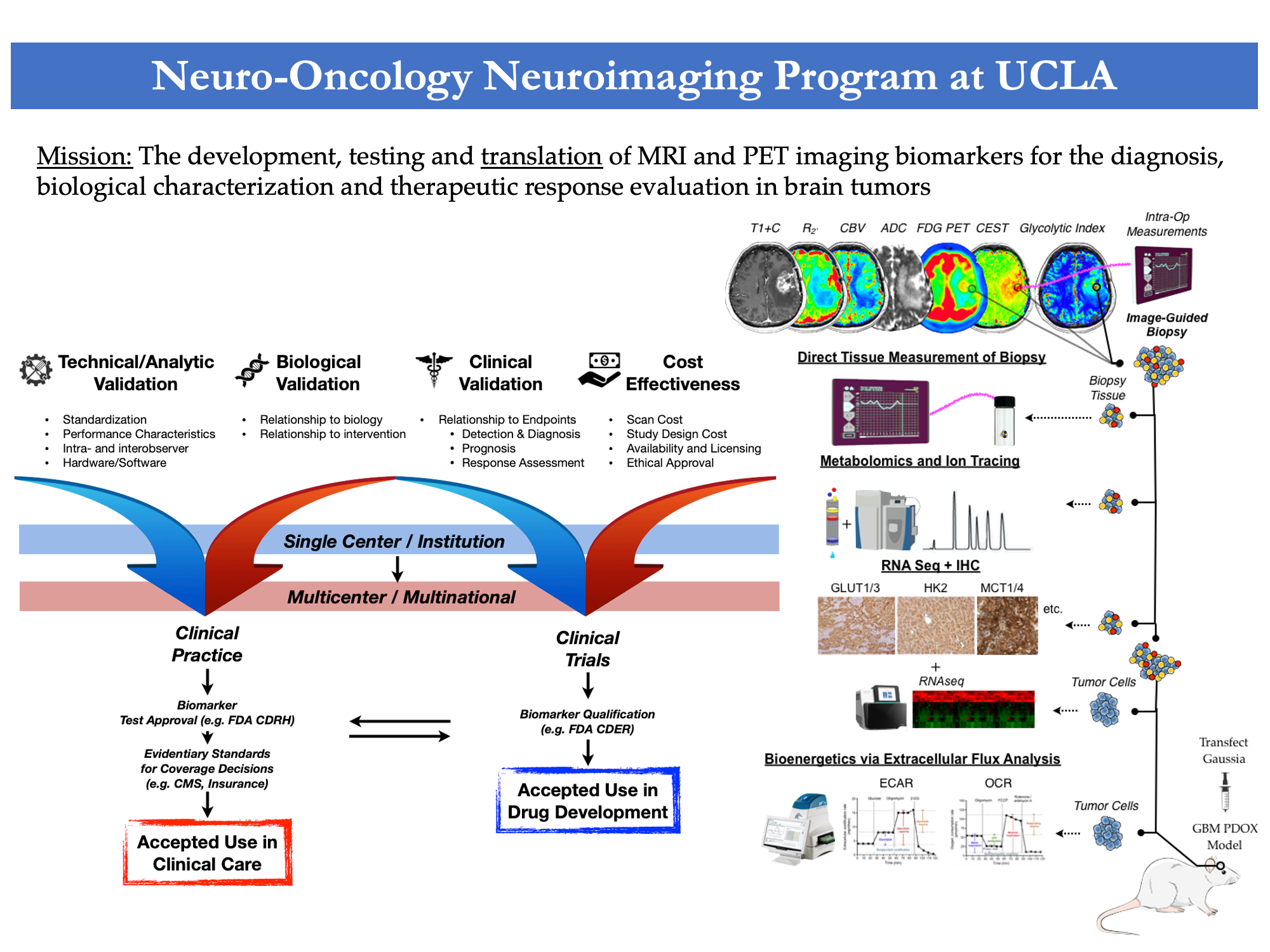
Our clinical research is dedicated to unraveling the complexities of brain tumors, encompassing a range of types from gliomas to metastatic lesions centering around the exploration and application of advanced MRI (Magnetic Resonance Imaging) and PET (Positron Emission Tomography) techniques for a comprehensive understanding of brain tumors. Through advanced MRI and PET techniques, researchers aim to:
-
Treatment Evaluation: BTIL is committed to assessing the effectiveness of brain tumor treatments. By utilizing advanced imaging, researchers can track changes in tumor size, composition, and metabolic activity, providing vital insights into treatment response and guiding clinical decisions.
-
Characterization: The lab specializes in decoding the complex makeup of brain tumors. By employing sophisticated imaging techniques, researchers can discern unique tumor characteristics, such as genetic markers, cellular density, and molecular signatures, aiding in accurate diagnosis and tailored treatment strategies.
-
Visualization: Advanced imaging enables BTIL to create detailed visual representations of brain tumors. These visuals enhance the understanding of tumor location, size, and interaction with surrounding brain structures, assisting surgeons and oncologists in planning precise interventions.
-
Quantification: Quantitative analysis is a cornerstone of BTIL's research. Through precise measurement and quantification of tumor features, such as blood flow, diffusion, and metabolic activity, researchers can extract quantitative data to inform clinical decisions and monitor treatment progress.
Our goal is to contribute to the advancement of brain tumor research and patient care. The lab's dedication to refining imaging technologies leads to improved diagnostic accuracy, personalized treatment strategies, and enhanced outcomes for individuals affected by brain tumors.
Selected References
1. |
Imaging biomarkers for antiangiogenic therapy in malignant gliomas.
Leu K, Pope WB, Cloughesy TF, Lai A, Nghiemphu PL, Chen W, Liau LM, Ellingson BM.CNS Oncol. 2013 Jan;2(1):33-47. doi: 10.2217/cns.12.29. |
2. |
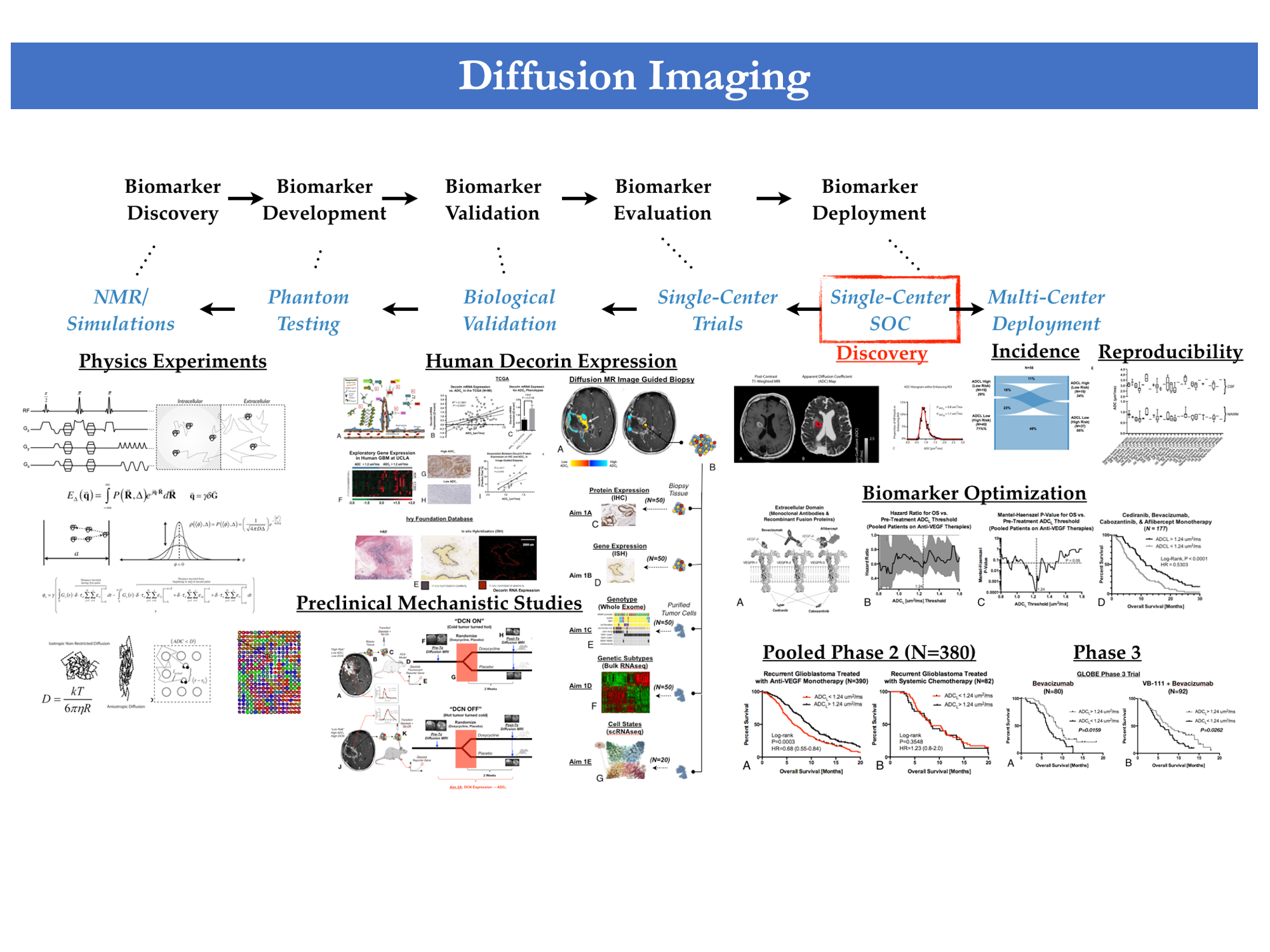
Validation of diffusion MRI as a biomarker for efficacy using randomized phase III trial of bevacizumab with or without VB-111 in recurrent glioblastoma.
Ellingson BM, Patel K, Wang C, Raymond C, Brenner A, de Groot JF, Butowski NA, Zach L, Campian JL, Schlossman J, Rizvi S, Cohen YC, Lowenton-Spier N, Minei TR, Shmueli SF, Wen PY, Cloughesy TF. Neurooncol Adv. 2021 Jun 19;3(1):vdab082. doi: 10.1093/noajnl/vdab082. |
3. |
Diffusion MRI Phenotypes Predict Overall Survival Benefit from Anti-VEGF Monotherapy in Recurrent Glioblastoma: Converging Evidence from Phase II Trials.
Ellingson BM, Gerstner ER, Smits M, Huang RY, Colen R, Abrey LE, Aftab DT, Schwab GM, Hessel C, Harris RJ, Chakhoyan A, Gahrmann R, Pope WB, Leu K, Raymond C, Woodworth DC, de Groot J, Wen PY, Batchelor TT, van den Bent MJ, Cloughesy TF. Clin Cancer Res. 2017 Oct 1;23(19):5745-5756. doi: 10.1158/1078-0432.CCR-16-2844. |
Neurological, neurodegenerative, and neuropsychiatric diseases
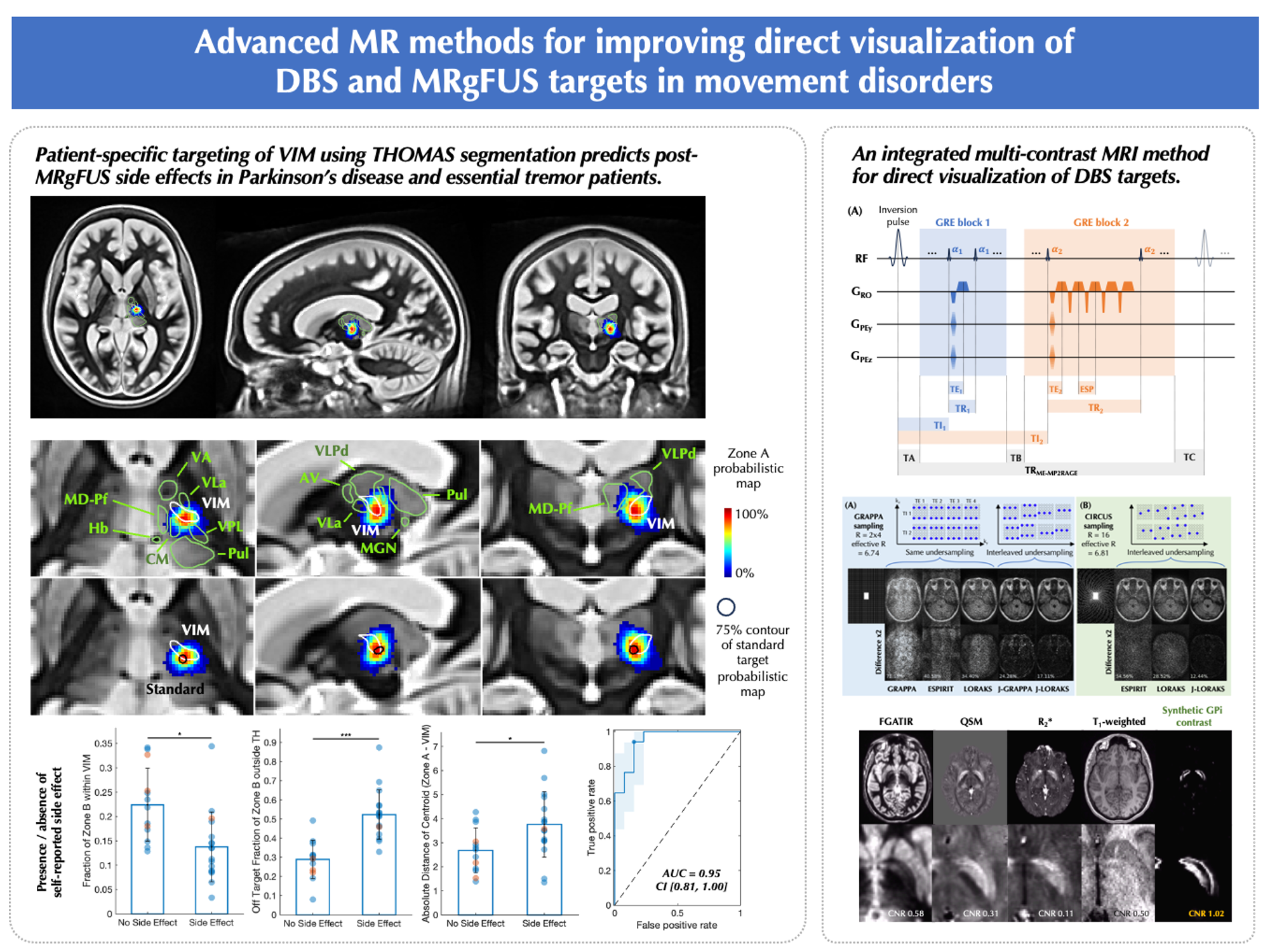
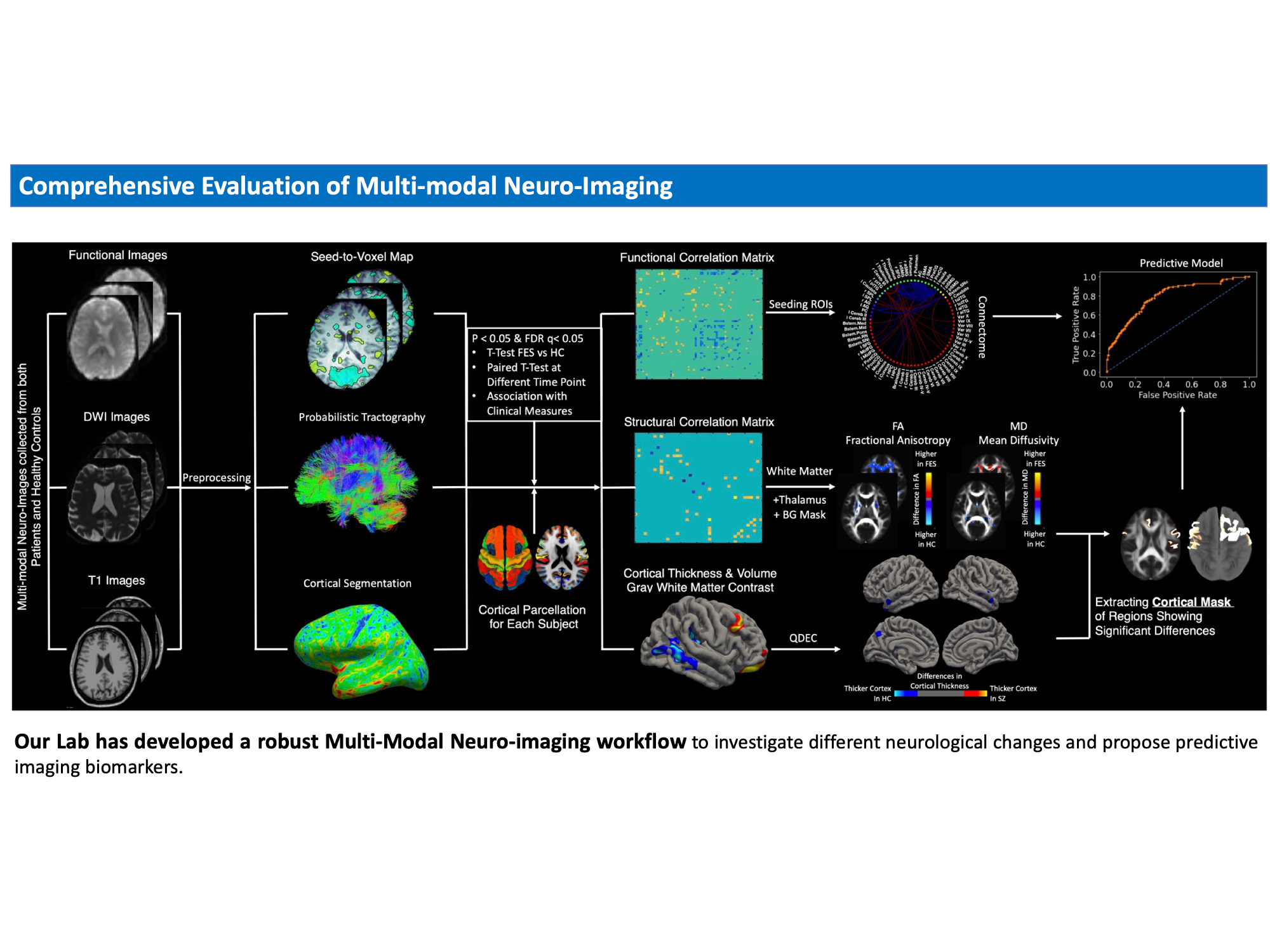
UCLA Brain Tumor Imaging Laboratory (BTIL) specializes in multimodal image processing and analysis, particularly concerning neurodegenerative diseases.
This comprehensive approach enables BTIL to provide a more holistic understanding of neurodegenerative diseases, such as Alzheimer's, Parkinson's, or Huntington's disease and CSM, Schizophrenia, and Epilepsy. By deciphering the combined information from multiple imaging modalities, BTIL contributes to improved disease diagnosis, progression tracking, and treatment evaluation, ultimately leading to better insights into disease mechanisms and more effective strategies for early detection and intervention.
Selected References
1. |
Diffusion MRI Phenotypes Predict Overall Survival Benefit from Anti-VEGF Monotherapy in Recurrent Glioblastoma: Converging Evidence from Phase II Trials.
Ellingson BM, Gerstner ER, Smits M, Huang RY, Colen R, Abrey LE, Aftab DT, Schwab GM, Hessel C, Harris RJ, Chakhoyan A, Gahrmann R, Pope WB, Leu K, Raymond C, Woodworth DC, de Groot J, Wen PY, Batchelor TT, van den Bent MJ, Cloughesy TF. Clin Cancer Res. 2017 Oct 1;23(19):5745-5756. doi: 10.1158/1078-0432.CCR-16-2844. |
2. |
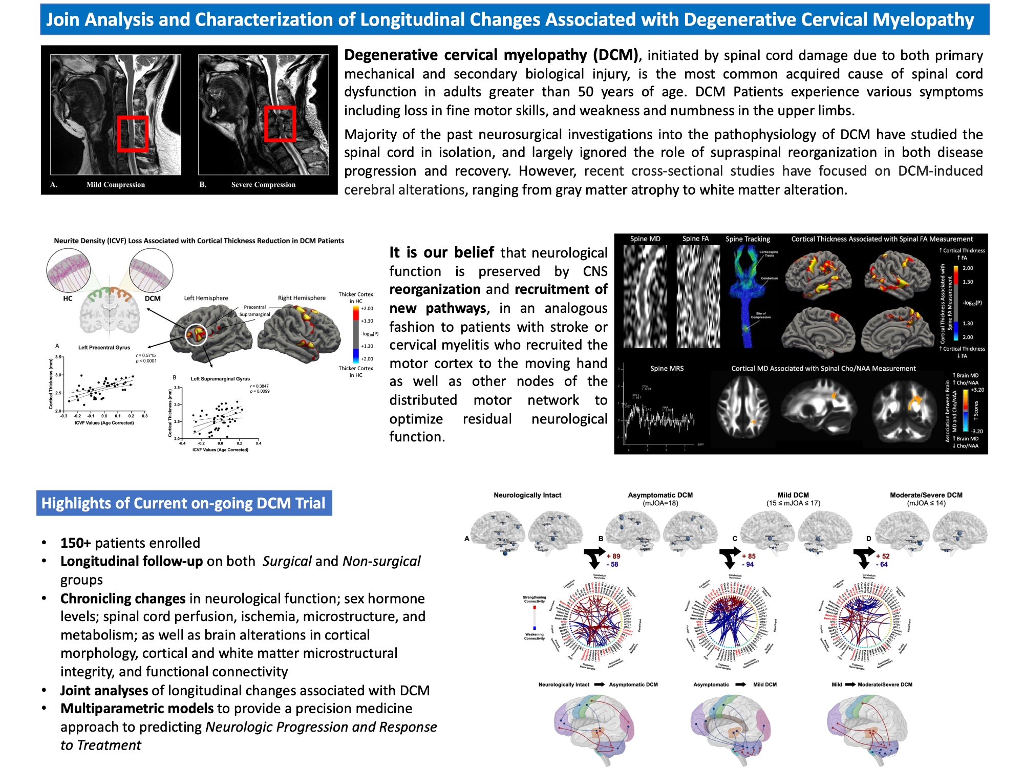
Evolution of brain functional plasticity associated with increasing symptom severity in degenerative cervical myelopathy.
Wang C, Ellingson BM, Oughourlian TC, Salamon N, Holly LT. EBioMedicine. 2022 Oct;84:104255. doi: 10.1016/j.ebiom.2022.104255. |
3. |
Cortical morphometric correlational networks associated with cognitive deficits in first episode schizophrenia.
Wang C, Oughourlian T, Tishler TA, Anwar F, Raymond C, Pham AD, Perschon A, Villablanca JP, Ventura J, Subotnik KL, Nuechterlein KH, Ellingson BM. Schizophr Res. 2021 May;231:179-188. doi: 10.1016/j.schres.2021.04.001. |
4. |
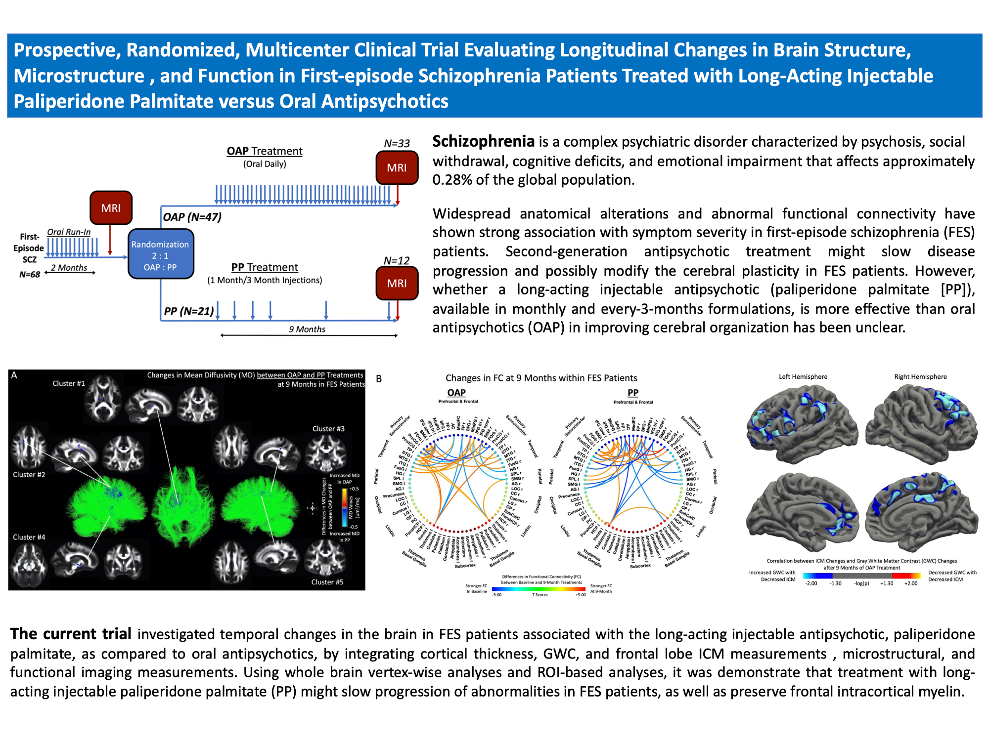
Cortical thickness, gray-white matter contrast, and intracortical myelin in first-episode schizophrenia patients treated with long-acting paliperidone palmitate versus oral antipsychotics.
Wang C, Tishler TA, Nuechterlein KH, Ellingson BM. Psychiatry Res. 2023 Aug;326:115364. doi: 10.1016/j.psychres.2023.115364. |
Imaging
Physics
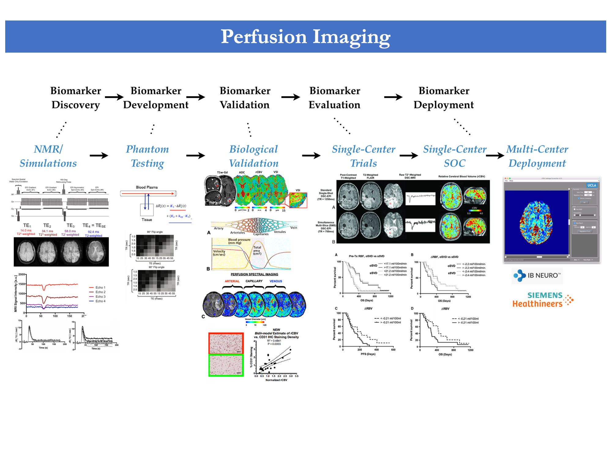
In collaboration with Siemens Healthineers and members of the UCLA MRRL, advanced MRI imaging techniques are continuously implemented at the BTIL with the goal of developing novel imaging contrasts and biomarkers to improve the characterization of malignant brain lesions and tumors, both for clinical research and diagnostic applications. To improve patient comfort and enrich the value of clinical exams, a second focus lies in reducing total image acquisition time by applying multi-slice and multinuclear interleaving acquisition techniques. Our endeavor of incorporating state-of-the-art techniques into our workflow will further our mission of pioneering diagnosis for brain malignancies and tumors.
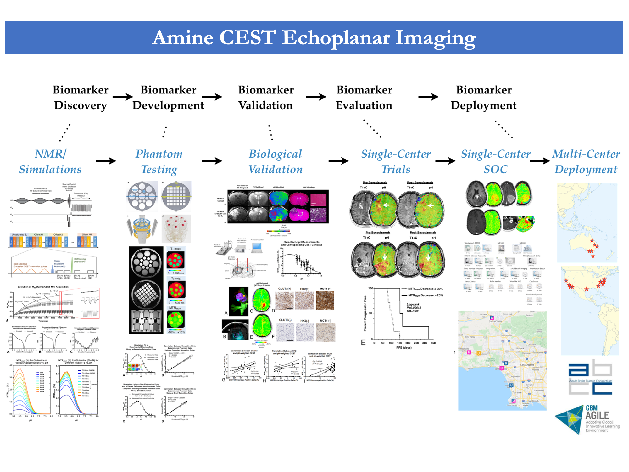
Chemical Exchange Saturation Transfer (CEST)
Our research delves into the domain of Chemical Exchange Saturation Transfer (CEST) sequences, with the aim of enhancing the sensitivity and specificity of imaging certain molecules. This novel approach contributes to the detailed characterization of brain tumors and their microenvironment.
Selected References
1. |
|
2. |
|
3. |
|
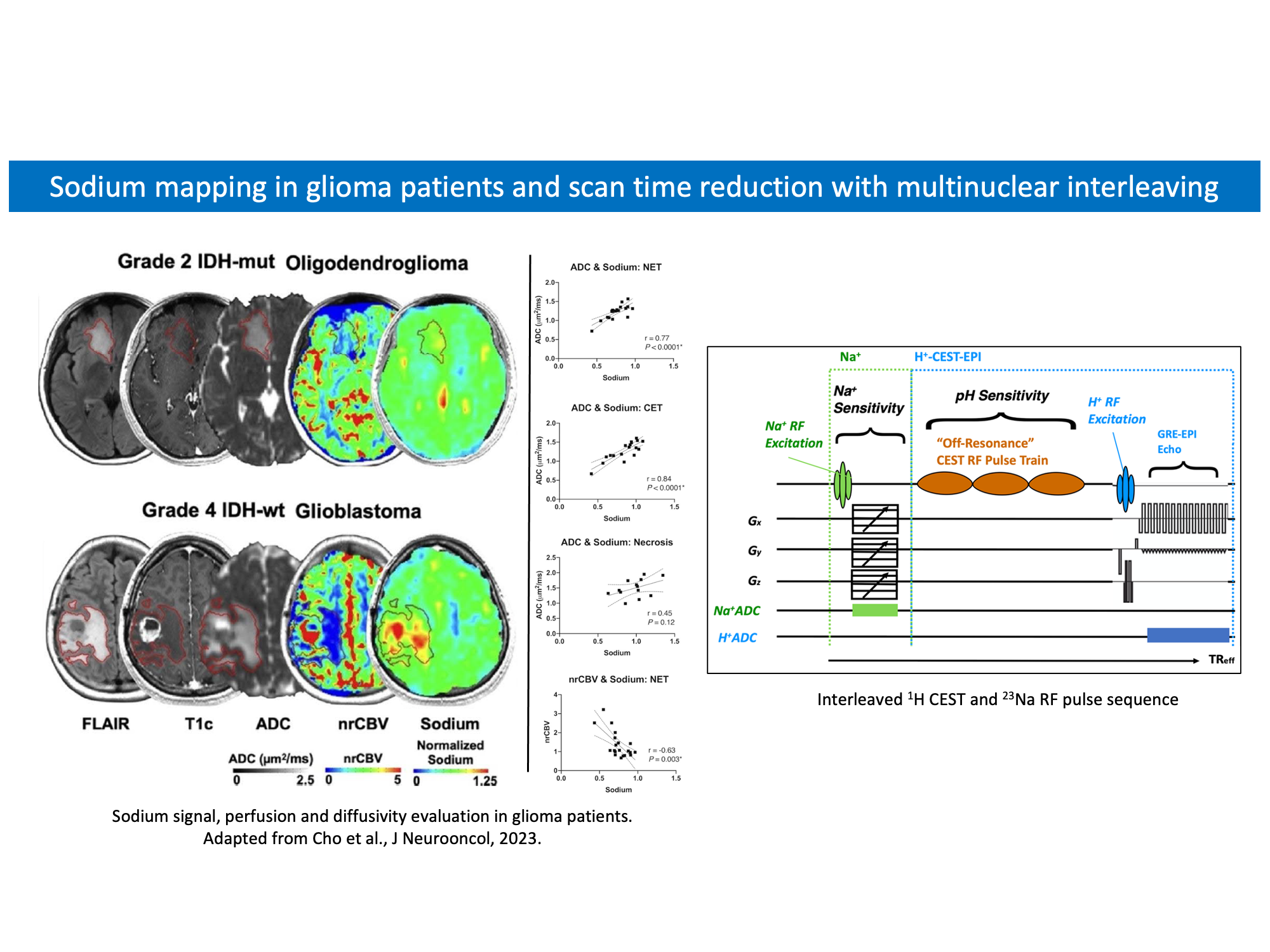
Sodium imaging
Evaluation of brain total sodium content offers unique insights into cellular integrity and viability. Although challenging to perform at 3T, the sodium gradient between extracellular and intracellular spaces makes it an excellent probe for Na+/K+-ATPase activity disruption. Sodium imaging therefore offers a great potential for evaluating brain diseases and tumor therapeutic response.
Selected References
1. |
|
2. |
|
3. |
|
Computational
Research
Deep Learning and Computer Vision in Medical Imaging
BTIL's expertise lies in leveraging advanced artificial intelligence (AI) technologies to enhance the analysis and interpretation of medical images. Our computational research branch leverages the capabilities of Deep Learning and Computer Vision techniques to unlock meaningful insights from complex medical imaging data.
By training these algorithms on vast datasets, BTIL aims to create automated tools that can assist in various aspects of medical imaging research:
-
Image Segmentation: Delineate regions of interest, enabling accurate identification of tumor boundaries and associated structures.
-
Feature Extraction: Extract intricate features, aiding in the identification of subtle patterns and disease markers.
-
Diagnostic Assistance: Detect anomalies, potentially leading to early and more accurate disease detection.
-
Generative Models: Synthetic data that simulates real medical images, aiding in the augmentation of limited datasets.
These technologies hold the potential to expedite analysis, improve accuracy, and unlock new layers of information within medical images, thereby advancing our understanding of brain tumors and paving the way for more effective diagnostic and treatment approaches.
Selected References
1. |
Synthesizing MR Image Contrast Enhancement Using 3D High-Resolution ConvNets. Chen C, Raymond C, Speier W, Jin X, Cloughesy TF, Enzmann D, Ellingson BM, Arnold CW. |
2. |
Federated learning enables big data for rare cancer boundary detection. Pati S, Baid U, Edwards B, Sheller M, ...., Ellingson BM, Cloughesy TF, Raymond C, Oughourlian T, Hagiwara A, Wang C, To MS, Bhardwaj S, Chong C, Agzarian M, Falcão AX, Martins SB, Teixeira BCA, Sprenger F, Menotti D, Lucio DR, LaMontagne P, Marcus D, Wiestler B, Kofler F, Ezhov I, Metz M, Jain R, Lee M, Lui YW, McKinley R, Slotboom J, Radojewski P, Meier R, Wiest R, Murcia D, Fu E, Haas R, T… |
Preclinical and
Cellular Imaging
The preclinical research arm of the UCLA Brain Tumor Imaging Laboratory (BTIL) delves into the foundational aspects of brain tumor research. Operating at the intersection of laboratory investigation and translational medicine, our preclinical studies involve the in-depth examination of brain tumor models in controlled environments. Through meticulous experimentation, data analysis, and the application of advanced imaging technologies, our goal is to uncover fundamental insights into the intricate mechanisms underlying brain tumors. These insights not only drive our clinical efforts but also pave the way for innovative therapeutic approaches in the battle against brain cancer.
Selected References
1. |
|
2. |
|
3. |
|